Microscopes are indispensable tools in science, allowing us to observe the microscopic world invisible to the naked eye. From biology and medicine to materials science and forensics, microscopes unlock a universe of detail. But simply having a microscope isn’t enough. Understanding its parts and how to use it correctly is crucial for obtaining clear images and accurate data. That’s where a Microscope Parts and Use Worksheet comes in handy! This worksheet serves as a practical guide, helping students and professionals alike familiarize themselves with the different components of a microscope and master the proper techniques for using it.
A well-designed worksheet typically covers several key areas. First, it introduces the various parts of the microscope, often including a labeled diagram. Students are asked to identify these parts and describe their functions. Secondly, it explains the steps involved in preparing a specimen for viewing. This might include learning how to create wet mounts or staining techniques. Finally, the worksheet guides users through the process of focusing, adjusting the light, and calculating magnification. By working through a comprehensive worksheet, users can develop a strong foundation in microscopy.
Mastering the microscope is not just about memorizing names and functions; it’s about developing a skill set. The worksheet fosters critical thinking by prompting users to consider the impact of different adjustments on the image quality. For example, understanding how to adjust the condenser or the diaphragm can dramatically improve contrast and resolution. Moreover, proper microscope usage is essential for accurate observation and interpretation of data. An ill-prepared slide or a poorly focused image can lead to incorrect conclusions and flawed research. Therefore, investing the time to learn the correct techniques is a worthwhile endeavor for anyone working with microscopes.
Using a Microscope Parts and Use Worksheet offers a structured and effective way to learn microscopy. It provides a hands-on learning experience that reinforces theoretical knowledge. Whether you’re a student learning biology, a medical technician analyzing blood samples, or a researcher studying cells, a solid understanding of microscope parts and use is essential. So, grab a worksheet, grab a microscope, and get ready to explore the microscopic world!
Microscope Parts and Use Worksheet: Answers
Below is a sample answer key, presented in HTML list format, for a hypothetical Microscope Parts and Use Worksheet. Remember that the specific questions and answers will vary depending on the worksheet.
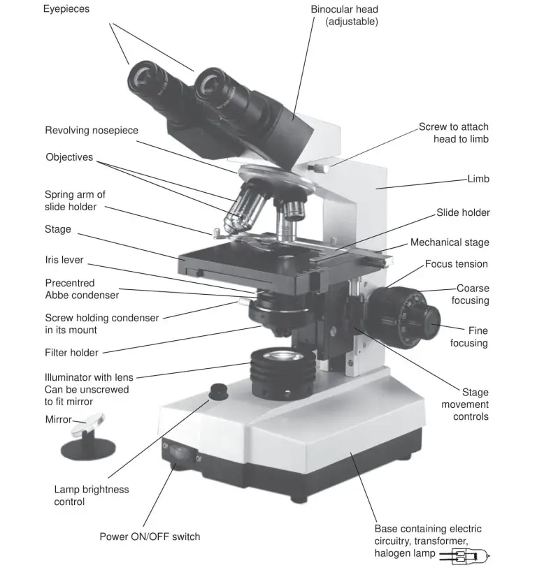
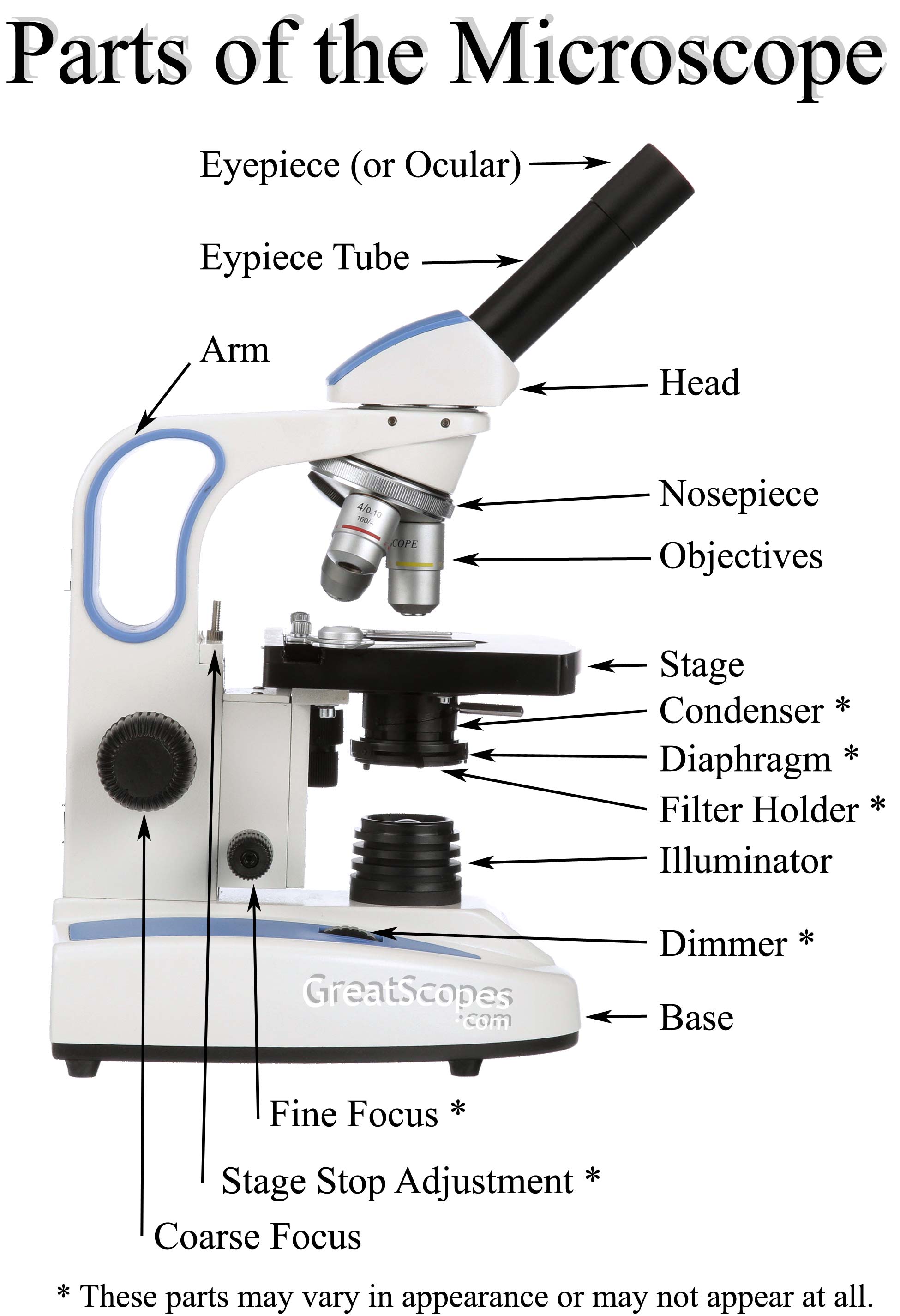
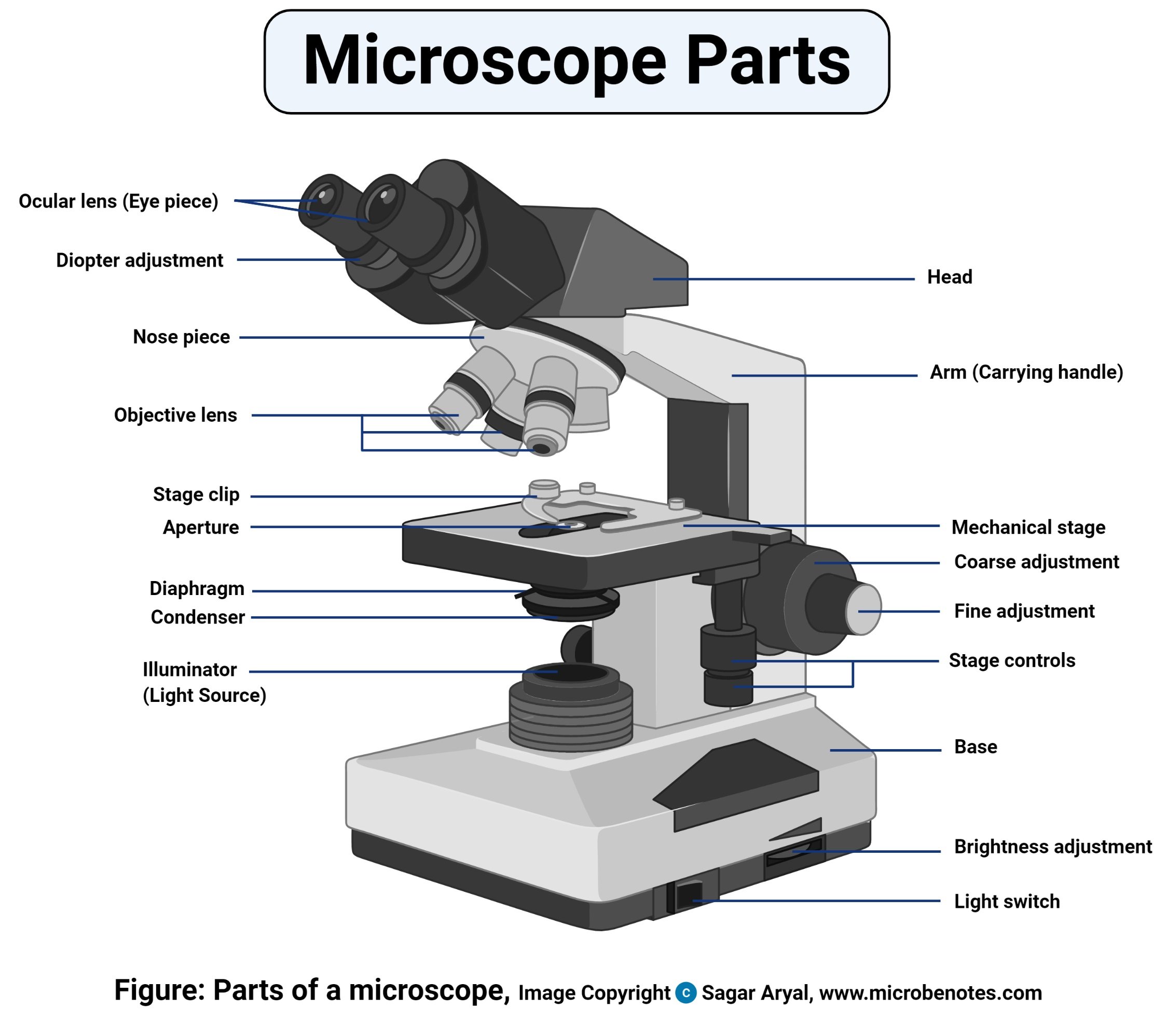
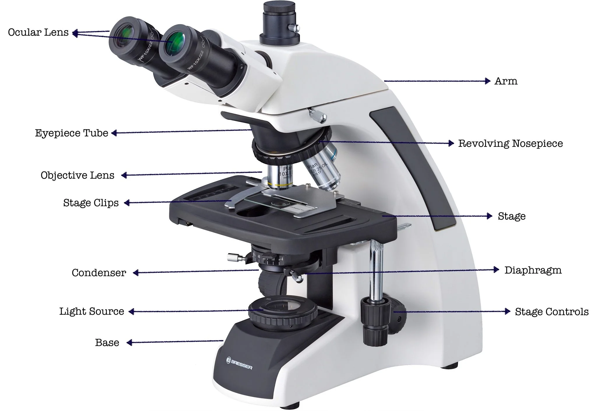
Part 1: Microscope Parts Identification
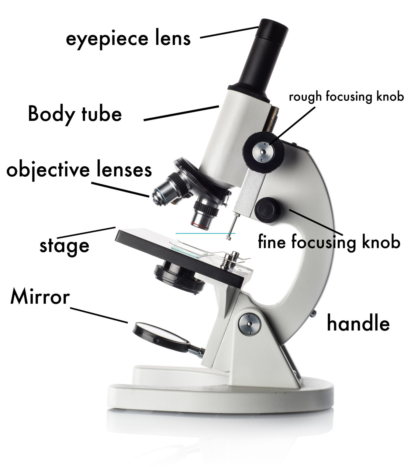
- Eyepiece (Ocular Lens): The lens you look through to view the specimen; typically magnifies 10x.
- Objective Lenses: Lenses with different magnifications (e.g., 4x, 10x, 40x, 100x) used to magnify the specimen.
- Nosepiece: Rotates to switch between different objective lenses.
- Stage: The platform where the specimen slide is placed.
- Stage Clips: Hold the specimen slide in place on the stage.
- Aperture: Hole in the stage that allows light to pass through the specimen.
- Illuminator: The light source that provides light for viewing the specimen.
- Condenser: Focuses the light onto the specimen, improving image contrast and resolution.
- Diaphragm (Iris): Controls the amount of light passing through the specimen. Adjusting the diaphragm affects contrast and depth of field.
- Coarse Adjustment Knob: Used for initial focusing, especially with low-power objectives. Moves the stage or nosepiece significantly.
- Fine Adjustment Knob: Used for fine-tuning the focus after using the coarse adjustment knob. Moves the stage or nosepiece very slightly.
- Base: The bottom support of the microscope.
- Arm: Supports the body tube and is used to carry the microscope.
Part 2: Specimen Preparation
- Wet Mount Preparation: Place a small sample on a clean slide, add a drop of water or appropriate solution, and cover with a coverslip. Avoid air bubbles.
- Staining (Example: Gram Staining): Follow the specific staining procedure for the desired stain. Staining enhances the visibility of certain structures. (Gram staining involves crystal violet, iodine, decolorizer, and safranin).
Part 3: Microscope Use and Calculations
- Steps for Focusing:
- Place the prepared slide on the stage and secure it with stage clips.
- Start with the lowest power objective lens (e.g., 4x).
- Use the coarse adjustment knob to bring the specimen into approximate focus.
- Use the fine adjustment knob to sharpen the focus.
- Adjust the light using the diaphragm and condenser.
- To view at higher magnification, carefully rotate the nosepiece to the next higher objective lens. Refocus using only the fine adjustment knob.
- Total Magnification Calculation: Multiply the magnification of the eyepiece lens by the magnification of the objective lens.
- Example: Eyepiece (10x) x Objective lens (40x) = Total Magnification (400x)
- Adjusting Light Intensity: Increasing the light intensity can improve visibility, but too much light can wash out the image. Adjust the diaphragm and illuminator to find the optimal balance for the specific specimen and objective lens.
This sample answer key should provide a general idea of the content covered in a typical Microscope Parts and Use Worksheet. Remember to consult your specific worksheet and resources for the correct answers and instructions. Good luck exploring the microscopic world!
If you are looking for What Is The Safest Way To Carry A Microscope you’ve came to the right page. We have 12 Images about What Is The Safest Way To Carry A Microscope like 19 Parts Of A Microscope And Their Functions – RankRed, Different types of microscopes used in microbiology ⋆ AgVetnepal and also Simple Microscope Diagram And Functions. Read more:
What Is The Safest Way To Carry A Microscope

studyritualizes.z4.web.core.windows.net
10 Parts Of A Microscope
mavink.com
Beginner's Guide To Buying A Microscope
learning-center.homesciencetools.com
Nome Das Partes De Um Microscópio – EDUCA
w20.b2m.cz
Microscope Photonique Monoculaire – UNITRADE
unitrade.sn
Parts Of A Microscope With Functions And Labeled Diagram
microbiologystudy.com
Microscope Summary | Britannica
www.britannica.com
Simple Microscope Diagram And Functions
ar.inspiredpencil.com
Microscope Optika (Italian) | Lab Supply

lab-supply.net
19 Parts Of A Microscope And Their Functions – RankRed
www.rankred.com
Simple Diagram Of Microscope
guidelibgatherings.z21.web.core.windows.net
Different Types Of Microscopes Used In Microbiology ⋆ AgVetnepal

agvetnepal.com
10 parts of a microscope. 19 parts of a microscope and their functions. Different types of microscopes used in microbiology ⋆ agvetnepal